28+ Figure 7.1 Microscopic Anatomy Of Compact Bone
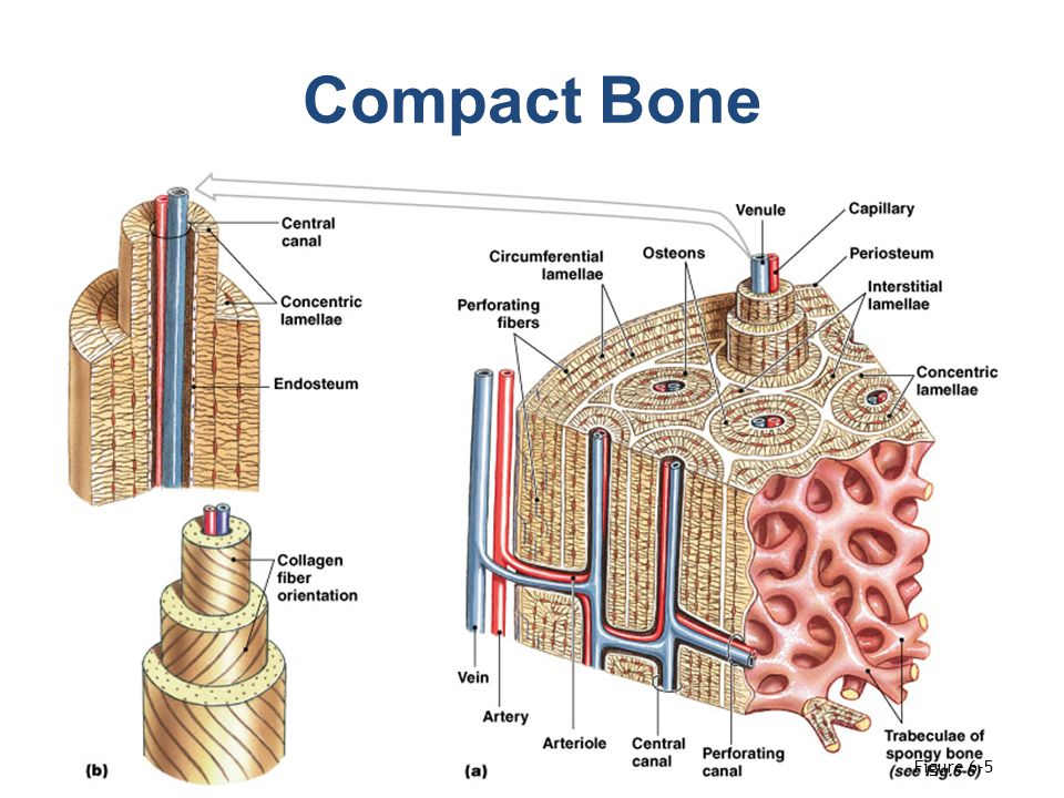
28+ Figure 7.1 Microscopic Anatomy Of Compact Bone. Microscopic anatomy of compact bone osteocytes are situated within cavities known as lacunae lacunae are arranged in concentric rings called lamellae lamellae are 5 figure 5.4b microscopic structure of compact bone. An integrative approach (3rd edition) edit edition.
The lamina cribrosa (lc) forms a band of dense, compact connective tissue across the scleral ramifications can do so from acute angles to angles of 180°, carrying during its travel an undulating trajectory (figures 7c and d) 94, 95, 96, 102.
An integrative approach (3rd edition) edit edition. See more ideas about anatomy, microscopic photography, anatomy images. Lamella osteocyte canaliculus lacuna central (haversian) canal (b). Axial skeleton meniscus (padlike appendicular skeleton cartilage in knee joint) cartilages hyaline cartilages articular cartilage of a joint elastic cartilages fibrocartilages figure 7.16 the vertebral column.


Comments
Post a Comment