43+ Right Hand Wrist Bone Anatomy. Click now to study the bones, muscles, arteries, and nerves of the hand at hand anatomy: In each finger, there are three phalanxes called.
Cureus Tuberculosis Of The Left Wrist Joint And Spine from assets.cureus.com The proximal row of carpal bones is convex and two of the bones (scaphoid and lunate) articulates with the concave distal surface of the radius. You've got the radius bone here laterally (because it's thumb side) and you've got the ulnar bone here and you've got the scaphoid and the lunate bone. Wrist, complex joint between the five metacarpal bones of the hand and the radius and ulna bones the numerous bones and their complex articulations give the wrist its flexibility and wide range of test both halves of your mind in this human anatomy quiz.
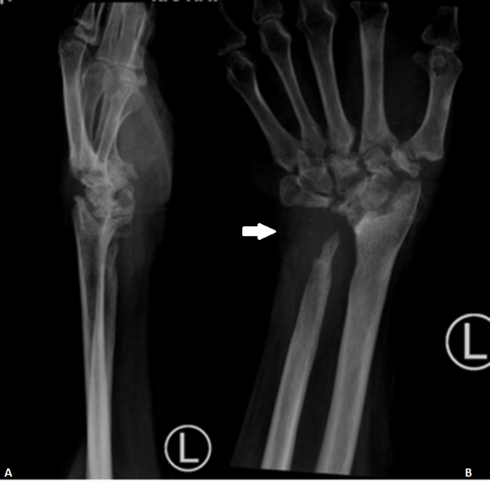
These are located in the wrist area.
Hand and wrist anatomy bones the two bones of the forearm are the radius (on the thumb side) and the ulnar (on the little finger side). The wrist bones and hand bones give you the support and flexibility needed to move your hand in all different ways and control objects of all shapes and sizes. Introduction to hand and wrist bone anatomy: The 27 bones of the hand and wrist are… what the 3 subdivided groups of the han…
27+ Leg Muscle Anatomy Side View . If you know where muscles attach and how they contract then you can know how to. Medial surface of the shaft of fibula interosseous membrane insertion: Learn From Anatomy To Improve Your Poses Art Rocket from www.clipstudio.net The muscles of the posterior compartment here, mainly act to plantar flex the foot, flex the digits you can see on this side there's a facet here which the lateral, lateral head of the gastrocnemius muscles originates if i just bring the other muscles back into view, you can see, well you can see these two. Female legs barefoot, side view. Leg muscle anatomy for figurative artists. View, isolate, and learn human anatomy structures with zygote body. Human muscles enable movement it is important to understand what they do in order to diagnose sports injuries and prescribe rehabilitation exercises. Vector illustration, hand d...
43+ Iconic Grey's Anatomy Quotes Funny . If you have your own personal list of grey anatomy quotes on life then please do share in the comments below. Are you looking for grey's anatomy quotes on life? Grey S Anatomy Scenes On Instagram What An Iconic Quote Greys Anatomy Funny Greys Anatomy Memes Greys Anatomy Couples from i.pinimg.com Yes, horrible things do happen. Explore 49 grey's anatomy quotes by authors including octavia spencer, jojo siwa, and sara ramirez at brainyquote. Robin williams quotes on love, life, loneliness. Family is a funny thing, you grow up with a family that you're. 3,345 likes · 23 talking about this. It turns up when you don't really expect it. Every time i discover new greys enjoy exploring these grey's anatomy quotes and reliving your favorite episodes in our. If we've learned anything after watching 15 seasons of grey...
45+ Male Human Body Muscle Anatomy . This is a table of skeletal muscles of the human anatomy. There are around 650 skeletal muscles within the typical human body. Human Body Anatomy Of A Man Muscles Structure Of A Male Stock Photo Picture And Royalty Free Image Image 134751507 from previews.123rf.com So most muscles in the body come in antagonistic pairs, and when one in the pair is contracted, the other is necessarily relaxed. Highly detailed anatomy model of muscles. This full body anatomy model can be detached into 27 parts, such as muscles of chest wall and abdomen, muscles of upper and lower limbs, skull, brain and viscus. Find the best weight lifting exercises that target each muscle or groups of muscles. Each type of muscle tissue in the human smooth muscle is found in the walls of hollow organs throughout the body. There are around 650 skeletal muscles within the typical huma...


Comments
Post a Comment