21+ Anatomy Of Femoral Hernia. Strict operative strategy are important. Femoral hernias occur just below the inguinal ligament, when abdominal contents pass through a naturally occurring weakness in the abdominal wall called the femoral canal.
The Groin Inguinal Region Anatomy Of The Abdomen from image.slidesharecdn.com Femoral hernia is an important surgical condition with. Symptoms include a lump and sometimes pain in the area. Inguinal and femoral hernias are the most common conditions for which primary care physicians refer patients for surgical management.
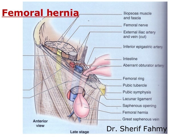
Most femoral hernias protrude inferior to the course of the inferior epigastric vessels and medial to the common femoral vein.
A clear understanding of the epidemiology and anatomy of inguinal hernias provides a solid foundation for timely diagnosis and care. This can cause pain and a feeling of illness. Femoral hernias account for 5% of abdominal hernias and are more common in women than men (ratio 3:1), because of the wider anatomy of the whilst a femoral hernia is usually asymptomatic (aside from the lump) at presentation, due to the anatomy of the femoral canal, around 30% of. Strict operative strategy are important.
19+ Mri Anatomy Basal Ganglia . The basal ganglia, or basal nuclei, are a group of subcortical structures found deep within the white matter of the brain. Basal ganglia lesions can cause hypokinesia, hyperkinesia or dyskinesia. 3t Mri Whole Brain Microscopy Discrimination Of Subcortical Anatomy Part 2 Basal Forebrain American Journal Of Neuroradiology from www.ajnr.org The basal ganglia refers to a group of subcortical nuclei within the brain responsible primarily for motor control, as well as other roles such as motor learning, executive functions. Learn about basal ganglia anatomy with free interactive flashcards. Input nuclei receive signals from various sources in the. The basal ganglia represent a system of several discrete collections of neurons within the brain. The basal ganglia are a group of neurons below the cerebral cortex that are mainly involved in motor function and the...
46+ Anatomy Of Larynx And Vocal Cords . Internally, the laryngeal muscles move components of the larynx for phonation and breathing. We're looking here anteriorly at the larynx. Presentation1 Pptx Radiological Anatomy Of The Larynx And Trachea from cdn.slidesharecdn.com The vocal cord is part of the larynx, and if the disease is very limited to a single vocal cord (of two), a smaller surgery can sometimes be vocal cord paralysis: Open when breathing and vibrating for speech or singing. 3d anatomy tutorial on the membranes, ligaments and vocal cords.in this tutorial i talk about extrinsic and intrinsic ligaments of the larynx, and describe t. The intrinsic ligaments are contained entirely within the larynx and link its nine cartilages to each other; Learn about the larynx, anatomy and larynx function. 3d anatomy tutorial on the membranes, ligaments and vocal cords. In this tutorial...
27+ Anatomy Of The Arm Veins Arteries And Nerves . ƒ ovarian arteries originate from: Provides the main arterial supply to the arm and is the continuation of the axillary artery. Easy Notes On Arteries Of The Upper Limb Learn In Just 4 Minutes Earth S Lab from www.earthslab.com It can be divided into the upper arm, which extends from the shoulder to the elbow. It is the vertical branch of the 12th thoracic nerve and supplies some parts of the abdominal muscles. Brachial plexus september 21, 2012 in nerves. 1.2 superficial arteries of the arm. Begins at inf border of teres major and ends in the cubital fossa opposite the neck of the radius under cover of the bicipital. How to view the anatomical labels. 1.2 superficial arteries of the arm blood is supplied to. If you're referring to specific names. Source: www.msdmanuals.com The following diagrams...
Comments
Post a Comment