48+ Anatomy Of Eye Conjunctiva. Substantia propria is more uniform than in bulbar conjunctiva and is more closely attached to tarsus (so invasive lesions appear clinically flat), has arterioles, veins and complex capillary network, myelinated and nonmyelinated branches of trigeminal. The conjunctiva is the clear, thin membrane that covers part of the front surface of the eye and the inner surface of the eyelids.
Causes Complications And Treatment Of A Red Eye Bpj Issue 54 from bpac.org.nz The eye is a very delicate structure, eye anatomy is very complex if there is any problem occur in any part of the eye following diseases can occur Start studying anatomy of the eye. The eye is a paired organ, the organ of vision.
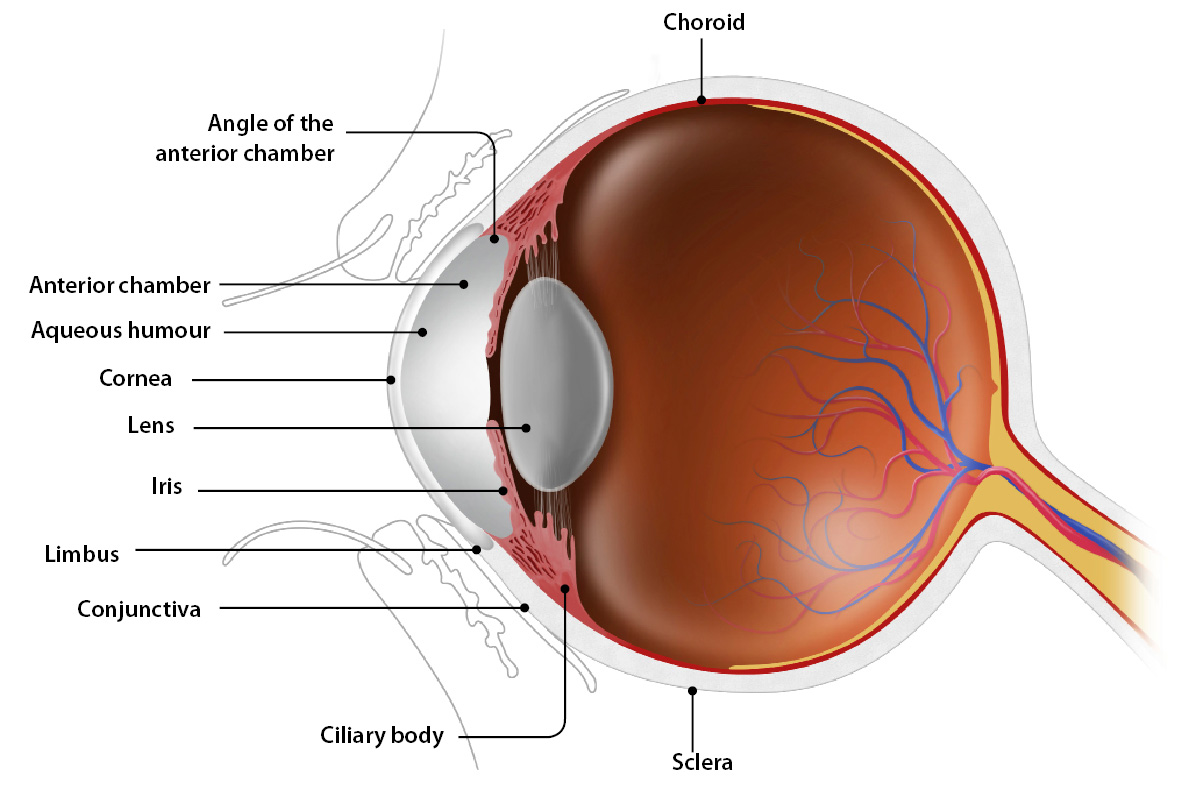
The clear tissue covering the white part of your eye and the inside of your eyelids.
The conjunctiva keeps bacteria and foreign material from getting behind the eye. The corneal epithelium (epithelium corneæ anterior layer) covers the front of the cornea and consists of several layers of cells. Pink eye, also called conjunctivitis, is a infection of the eye's conjunctiva usually caused by a bacteria or virus that results in red, itchy, painful eyes. It then flows back behind the eye, loops forward, and forms the inside.
27+ Leg Muscle Anatomy Side View . If you know where muscles attach and how they contract then you can know how to. Medial surface of the shaft of fibula interosseous membrane insertion: Learn From Anatomy To Improve Your Poses Art Rocket from www.clipstudio.net The muscles of the posterior compartment here, mainly act to plantar flex the foot, flex the digits you can see on this side there's a facet here which the lateral, lateral head of the gastrocnemius muscles originates if i just bring the other muscles back into view, you can see, well you can see these two. Female legs barefoot, side view. Leg muscle anatomy for figurative artists. View, isolate, and learn human anatomy structures with zygote body. Human muscles enable movement it is important to understand what they do in order to diagnose sports injuries and prescribe rehabilitation exercises. Vector illustration, hand d...
43+ Iconic Grey's Anatomy Quotes Funny . If you have your own personal list of grey anatomy quotes on life then please do share in the comments below. Are you looking for grey's anatomy quotes on life? Grey S Anatomy Scenes On Instagram What An Iconic Quote Greys Anatomy Funny Greys Anatomy Memes Greys Anatomy Couples from i.pinimg.com Yes, horrible things do happen. Explore 49 grey's anatomy quotes by authors including octavia spencer, jojo siwa, and sara ramirez at brainyquote. Robin williams quotes on love, life, loneliness. Family is a funny thing, you grow up with a family that you're. 3,345 likes · 23 talking about this. It turns up when you don't really expect it. Every time i discover new greys enjoy exploring these grey's anatomy quotes and reliving your favorite episodes in our. If we've learned anything after watching 15 seasons of grey...
45+ Male Human Body Muscle Anatomy . This is a table of skeletal muscles of the human anatomy. There are around 650 skeletal muscles within the typical human body. Human Body Anatomy Of A Man Muscles Structure Of A Male Stock Photo Picture And Royalty Free Image Image 134751507 from previews.123rf.com So most muscles in the body come in antagonistic pairs, and when one in the pair is contracted, the other is necessarily relaxed. Highly detailed anatomy model of muscles. This full body anatomy model can be detached into 27 parts, such as muscles of chest wall and abdomen, muscles of upper and lower limbs, skull, brain and viscus. Find the best weight lifting exercises that target each muscle or groups of muscles. Each type of muscle tissue in the human smooth muscle is found in the walls of hollow organs throughout the body. There are around 650 skeletal muscles within the typical huma...


Comments
Post a Comment