14+ Knee Anatomy Acl Pcl
14+ Knee Anatomy Acl Pcl. The acl and pcl are two major ligaments that crisscross within the joint, allowing the knee to flex and extend without sliding back and forth. Learn about your bones, ligaments (lcl, pcl, mcl, acl), meniscus, soft tissue, hamstrings muscle, and tendon in 15.
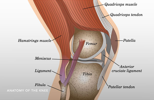
The anterior cruciate ligament (acl) and posterior cruciate ligament (pcl) form an each part of the anatomy needs to function properly for the knee to work.
For more anatomy content please follow us and visit our. Secondary restraint to tibial external rotation. What ligaments support the knee? The anterior cruciate ligament (acl) and posterior cruciate ligament (pcl) form an each part of the anatomy needs to function properly for the knee to work.


Comments
Post a Comment